
Cornea DSAEK
Advanced corneal transplant surgery (DSAEK) for corneal disorders - restoring vision with precision, safety and faster recovery.
Overview & Clinical Approach
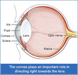
The human eye consists of three layers - sclera, choroid and retina. The front portion of the sclera is called the cornea, a thin, transparent tissue that covers the front of the eye and is the surface on which contact lenses sit. The cornea plays a major role in bending (refracting) light and is responsible for nearly two-thirds of the eye's focusing power, so it must remain smooth, compact and transparent for clear vision.
A modern, minimally invasive corneal transplant that replaces only the damaged inner layer of the cornea.
Research studies in India show that corneal blindness is one of the leading causes of blindness, with more than 10 lakh people blind in both eyes. Corneal blindness can often be restored with a corneal transplant.
Healthy donor corneas, donated soon after death, are transplanted to replace the damaged cornea. As noted by Dr. Jugal Shah, corneal transplants are highly successful, but eye donation awareness remains low in the country.
Traditionally, full-thickness corneal transplants were performed. Today, DSAEK (Descemet’s Stripping Automated Endothelial Keratoplasty) allows selective replacement of only the damaged endothelial layer, preserving healthy corneal tissue.
DSAEK is an outpatient procedure; the entire surgery typically takes 45-60 minutes and no hospital stay is required.
Common Symptoms
- Swollen, scarred or cloudy cornea resulting in reduced or lost vision
- Blurred vision that gradually improves post-procedure
- Corneal endothelial dysfunction symptoms
Primary Causes & Risk Factors
- Fuchs’ endothelial dystrophy
- Posterior polymorphous membrane dystrophy
- Congenital hereditary endothelial dystrophy
- Bullous keratopathy
- Iridocorneal endothelial (ICE) syndrome
- Failed previous corneal transplants
Diagnostic Procedures
- Corneal Pachymetry and Endothelial Cell Density (Specular Microscopy)
- Slit-lamp examination to evaluate corneal clarity and Descemet's layer
Treatment Steps
Endothelial Layer Preparation
A thin donor graft containing healthy endothelial cells is micro-cut and prepared.
Descemet's Stripping
The diseased endothelial layer is removed through a micro-incision.
Tissue Insertion & Air Bubble Positioning
The donor graft is inserted and held in place against the patient's cornea using an air bubble.
Recovery & Post-Operative Care
Full recovery usually takes about three months; vision is initially blurred and gradually improves within one to two weeks. Patients are advised to lie flat on their back for the first few days to help the graft attach properly (typically 45 minutes out of every hour), use prescribed antibiotic and steroid eye drops regularly, avoid rubbing or bumping the eye, wear protective eyewear when required, and avoid swimming, diving and heavy lifting for some time.
Who is Eligible for Cornea DSAEK?
- DSAEK is suitable only when damage is limited to the inner endothelial layer
- Patients with deeper or full-thickness corneal damage may require a full corneal transplant instead
Risks of DSAEK / DMEK Surgery
- In rare cases, the graft may not fully attach, requiring a small procedure to insert an additional air bubble
- Serious complications such as infection, retinal detachment or haemorrhage are extremely rare
- Minor pupil shape or size changes may occasionally occur
Key Treatment Benefits
- Minimally invasive selective layer transplantation preserving healthy corneal tissue
- Smaller incision with fewer stitches compared to full-thickness transplants
- Faster visual rehabilitation and higher long-term success rates
Consultation for Cornea DSAEK
Fill out the details below. Our care team will call to confirm your preferred OPD slot.